Unit 9 – Transport (Exercise Questions)
ANSWERS THE FOLLOWING QUESTIONS.
Q.1. How would you relate the internal structure of rot with the uptake of water and salt?
Q.2. Define transpiration and relate it with cell surface and with stomatal opening and closing.
Q.3. How do different factors affect the rate of transpiration?
Q.4. Transpiration is necessary evil. Give comments
Q.5. Explain the movement of water in terms of transpiration pull.
Q.6. Describe the theory of pressure flow mechanism to explain the translocation of food in plants.
Q.7. List the function of the components of blood.
Q.8. How do we classify blood groups in terms of the ABO and the Rh blood group system?
Q.9. State the sign and symptoms, causes and treatments of leukemia and Thalassaemia.
Q.10. What four chambers make the human heart and how blood flows through these chambers?
Q.11. Compare the structure and function of an artery, a vein and a capillary.
Q.12. Draw diagram which can illustrate the origins, locations and targets areas of the main arteries in human blood circulatory system
Q.13. How would you differentiate between atherosclerosis and arteriosclerosis?
Q.14. State the causes, treatments and prevention of myocardial infarction.
SHORT QUESTIONS – TEXT EXERCISE
Q.1. What are lenticels and where are they fund in plant body?
Q.2. What is the role of potassium ions in the opening of stomata?
Q.3. Define the cohesion-tension theory
Q.4. What do you mean by sources and sinks according to the pressure flow mechanism?
Q.5. What are the two main types of white blood cell? How do they differ?
Q.6. You are pus at the site of infection on you skin. How is it formed?
Q.7. What role does the pericardial fluid play?
Q.8. Define the terms systole and diastole.
THE TERMS TO KNOW
Q.1. How would you relate the internal structure of rot with the uptake of water and salt?
Answer:
INTERNAL STRUCTURE:
The centre of the root in most of the cases is occupied by vascular tissue. The xylem composed of conducting elements, the Tracheids and vessels occupies the centre of the root is continuous with the xylem tissue in the
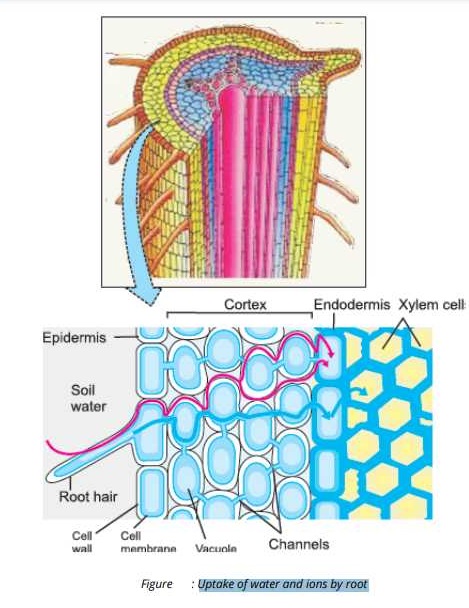
stem. The phloem tissue is closely associated to the xylem tissue. The xylem and phloem elements are surrounded by a layer of living cells, the Pericycle. The vascular tissue and the epicycle form a tube of conducting cells called stele. Just outside the stele is a layer of cells called endodermis. This endodermis acts as watertight jacket around the conducting vascular elements because water with its dissolved substances cannot pass around the endodermal cells via their walls.
Outside the endodermis, several layers of large thin walled living cells with intercellular spaces among them are present. This is dilled as cortex. The air spaces form interconnected air channels necessary for internal aeration. The cell wall of cortical cells are highly permeable to water and dissolved solutes. The cortex is surrounded by a layer of almost flattened cells. It is epidermis. Some epidermal cells develop long projections called Root hairs that extend out among the soil particles around the root. The root hairs increase soil root contact and enhance water absorption and the volume of soil penetrated.
| ylem .tissue is responsible for the transport of water and dissolved substances from roots to aerial parts. It consists of vessel elements and traccheids. Phloem tissue is responsible for the conduction of dissolved organic matter (food) between different parts. of plant body. It consists of sieve tube cells and companion cells. |
Water always moves from an area of higher water potential to an area of lower water potential. |
UPTAKE OF WATER AND SALTS
Root hairs provide large surface area for absorption. The cytoplasm of the root hairs has higher concentration of salts than the-soil water, so water moves by osmosis into the root hairs. Salts also enter root hairs, water and
salts must move through the epidermis and cortex of the root and then into.
the xylem tissue in the centre of the root.
After their entry into root hairs, water and salts travel through intercellular spaces or through cells (via channels, called plasmodesmata) and reach xylem tissue. Once in xylem, water and salts are carried to all the aerial parts of plant.
Q.2. Define transpiration and relate it with cell surface and with stomatal opening and closing.
Answer:
TRANSPIRATION:
Transpiration is the loss of water in the form of vapours from theaerial parts especially leaves.
TYPES OF TRANSPIRATION:
There are three types of transpiration:
(i) STOMATAL TRANSPIRATION
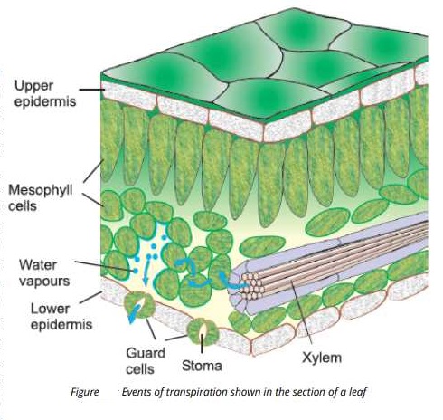
Evaporation of water through stomata is called stomatal transpiration. More than 90% of water is lost through the stomata although stomata openings surface. Stomatal transpiration involves two processes:
(a) Evaporation of water from cell wall surfaces bordering the inter cellular spaces, or air spaces of the mesophyll tissue.
(b) Diffusion of the water vapours from the intercellular spaces into the atmosphere by way of the stomata.
(ii) CUTICULAR TRANSPIRATION
The loss of water as it vapour, directly from the surfaces of leaves and herbaceous stems through the cuticle is called Cuticular Transpiration. Only a small fraction of water is lost by Cuticular Transpiration.
(iii) LENTICULAR TRANSPIRATION
The loss of water through the lenticels in the bark is called Lenticular Transpiration. Lenticels are small openings present in the bark.
Most plants keep their stomata open during the day and close them at night. The regulation of transpiration through stomata depends upon guard cells. Each stoma is surrounded by two guard cells, which are attached to each other at their ends. The inner concave sides of guard cells are thicker than the outer convex sides.
MECHANISM
Initially, it was thought that concentration of glucose in guard cells is responsible for opening and closing stomata. When guard cells become turgid, their shapes are like two beans and stoma between them opens. When the guard cells loose water and become flaccid, their inner sides touch each other and the stoma closes.
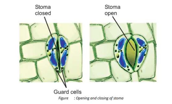
Recentally, it is has been revealed that opening and closing of stomata depends upon the movements of Potassium ions in and out of guard cells. The blue wavelength of daylight cause the K+ to flow into the guard cells,
from the surrounding epidermal cells. Water passively follows these ions into the guard cells. The guard cells become .turgid and open. During the nighttime, the K+ flows back to the surrounding epidermal cells, which also lead to loss of water. Guard cells become flaccid and stomata close.
Some plants open their stomata during night when overall water stress is low.
Q.3. How do different factors affect the rate of transpiration?
Answer:
Stomata movement i.e. opening and closing of stomata directly control the rate of transpiration while the stomatal movement is influenced by the light.
In strong light, the rate of transpiration is increased because the stomata are opened. While in dim light, the rate of transpiration is low because the stomata ‘are closed.
Following are the environmental factors affecting the rate of transpiration.
(i) Temperature’ (ii) Air Humidity
(iii) Air Movement (iv) Leaf Surface Area
(i) Temperature:
An increase in temperature causes an increase in the rate of transpiration. Because the high temperature is responsible for the reduction of the Humidity of the surrounding are and an increase in the
kinetic energy of water molecules.
The rate of transpiration increases with every rise of 10°C in temperature. However, if the temperature exceeds 40 – 45°C, the stomata are closed and the transpiration stops. As a result of which much of the needed water is not lost by the plant.
(ii) Air Humidity:
Dry air is responsible for increasing the rate of diffusion of water molecules from the surface of mesophyll cells, air spaces and through stomata to the outside of leaf. As a result of which more water is lost thus causing an increase in the rate of transpiration.
In Humid air the rate of diffusion of water molecules is decreased from the surface of the leaf; thus the rate of transpiration is low.
(iii) Air Movement:
The air in motion is called wind, which causes increase in the rate of diffusion of water molecules. As a result of which, the rate of evaporation of water from the surfaces of leaf mesophyll is faster through stomata.
(iv) Leaf Surface Area: The rate ‘of transpiration is directly proportional to the surface area of leaf. More surface area provides more stomata and the rate of transpiration is increased.
Q.4. Transpiration is necessary evil. Give comments.
Answer:
Transpiration has been described as necessary evil because it is potentially harmful process but is an invetiable too.
HARMFUL ASPECT
Transpiration is harmful in the sense that during the conditions of drought, loss of water from plant can lead to
wilting, serious dessication and often death of a plant if conditions of drought are experienced.
NECESSITY ASPECT
1. Transpiration creates a pulling. force called’ transpiration pull which is responsible for the conduction of water and mineral salts from the root, to the aerial parts of plant.
2. Evaporation of water from the exposed surface of cells of leaves by transpiration has a cooling effect on plant.
This cooling effect is particularly important in warmer environments.
3. Wet surface of leaf cells allow gaseous exchange between the plant and its environment, which is essential for photosynthesis and respiration.
Q.5. Explain the movement of water in terms of transpiration pull.
Answer:
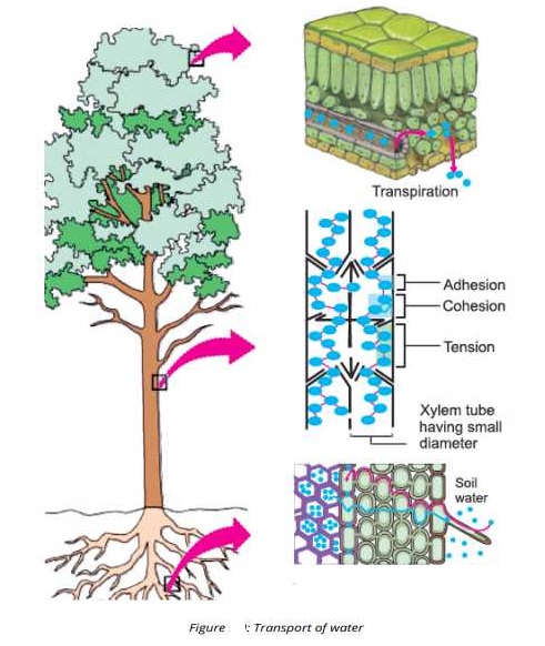
Transpiration pull is responsible for the upward movement of water. Water evaporates from the leaf surface’ during transpiration. As water evaporates from cells of the spongy and palisade layers of the leaf and passes out through the stomata, the concentration of solutes increases in the leaf cells. When this happens, water moves by osmosis into the leaf cells form xylem vessels and Tracheids of the leaf. As this water moves in cohesion between water molecules in the leaf cells and those in the xylem tissues makes possible the development of forces that pull water into the root up the stem, and out, into the leaves. This transpiration pull or cohesion and tension in water results in the upward movement of water from roots to stem and leaves.
Q.6. Describe the theory of pressure flow mechanism to explain the translocation of food in plants.
Answer:
This pulling force created by the transpiration of water is called transpiration pull. It also causes. water to move transversely (from root epidermis to cortex and Pericycle). Following are the reasons for the creation of transpiration pull.
• Water is held in a tube (xylem) that has small diameter.
• Water molecules adhere to the walls of xylem tube (adhesion).
• Water molecules cohere to each other (cohesion).
Q.7. List the function of the components of blood.
Answer:
Functions of Blood
Human blood performs the following functions:
(i) Transportation of martials
Blood transport the materials e.g. nutrients, water, salts and waste products to the different parts of body. Hormones are also transported by blood from the endocrine tissues to the target sites.
(ii) Transportation of gasses
Respiratory gases e.g. Oxygen and Carbon dioxide are transported by blood.
(iii) Protection from diseases
Blood helps in body’s defense against diseases.
(iv) Protection from microbes
Blood has particular proteins e.g. interferon (produced by liver) and antitoxins (produced by blood cells). Such chemicals protect the body from nucleic acids and toxins of invading organisms.
(v) Maintenance of pH
Blood acts as a buffer to maintain the acid-base balance i.e. concentration of hydrogen and hydroxyl in the body.
(vi) Maintenance of Homeostasis
Blood is responsible for maintaining the amounts of chemicals in the body to constant or nearly constant levels. So, blood also maintains homeostasis.
(vii) Exchange of materials
Blood helps in the exchange of materials between blood and body tissue through blood capillaries.
Q.8. How do we classify blood groups in terms of the ABO and the Rh blood group system?
Answer:
International Society of Blood Transfusion (ISBT) has recognized about 29 human blood group systems. These blood group systems are classified on the basis of presence or absence of antigens on the surface of red blood cells. The two of these blood group system are as follows:
(1) ABO Blood Group System
(2) Rh Blood Group System
1. ABO Blood Group System
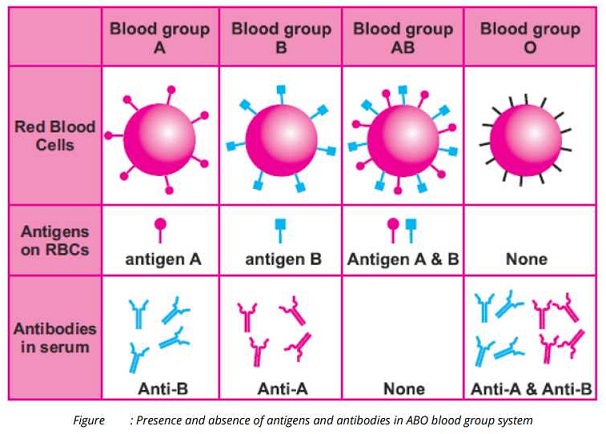
This system was discovered by the Austrian Scientist Karl Landsteiner in 1900. He has categorized blood into four groups on the presence or absence of specific antigens. Antigens A and B present on the surface of RBCs.
(i) Blood group A
A person having blood group A has antigen A on the red blood cells. This person contains anti-B antibodies is its blood serum.
(ii) Blood group B
A person having blood group B has antigen B on the red cells and because antigen A is absent so its blood serum contain anti-A antibodies.
(iii) Blood group AB
Both the antigens A and B will be present on the red blood cells and no any antibody will be present in the blood serum.
(iv) Blood group O
A person having none of the A and B antigens has blood group O and because both antigens are absent so both the anti-A and anti-B antibodies will be present.
Q.9. State the sign and symptoms, causes and treatments of leukemia and Thalassaemia.
Answer:
Leukaemia (Blood Cancer):
It is the production of large number of immature and abnormal white blood cells (leucocytes). This.is caused by a cancerous mutation in Bone marrow or Lymph tissue cells.
Treatment
It is a very serious disorder and the patient needs to change its blood regularly with the normal blood, got from donors. It can be cured by bone marrow transplant, which is in most cases effective, but very expensive
treatment.
Thalassaemia (g. thalassa = sea; haem = blood)
It is also called Cooley’s anemia on the name of Thomas B. Cooley, an American Pediatrician.
Causes:
It is genetically transmitted by the mutations in the gene of hemoglobin. The mutation results in the production of defective hemoglobin which does not transport oxygen properly.
Treatment:
The blood of these patients is to replace regularly with normal blood.
| The world celebrates the International Thalassaemia Day on If’ of May. This day is dedicated to raise public awareness about Thalassaemia and to highlight the importance of the care for Thalassaemia patients. |
There are about 60-80 million people in the world who carry Thalassaemia. India, Pakistan and Iran are seeing a large increase of Thalassaemia patients. Pakistan alone has 250,000 such patients. The patients require blood transfusions for lifetime. (Source: The Thalassaemia International Foundation) |
Q.10. What four chambers make the human heart and how blood flows through these chambers?
Answer:
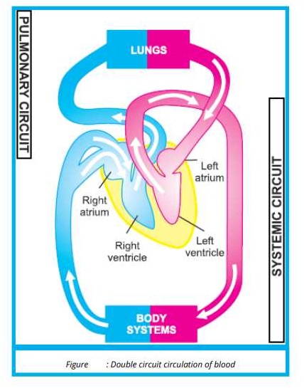
Pulmonary circulation / circuit
The pathway in which deoxygenated blood is carried from the heart to the lungs and in return oxygenated blood is carried from the lungs to the heart is called pulmonary circulation or circuit.
Systematic Circulation or circuit.
The pathway in which oxygenated blood is carried from the heart to the body tissues and in return deoxygenated blood is carried from the body tissues to the heart is called systemic circulation or circuit.
Q.11. Compare the structure and function of an artery, a vein and a capillary.
Answer:
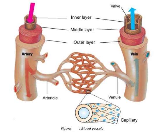
Blood circulatory system consists of blood vessels which are responsible for the transportation of blood throughout the body. Following are the important blood vessels of the circulatory system:
(i) Arteries (ii) Capillaries (iii) Veins
ARTERIES:
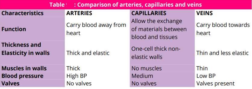
Arteries are the blood vessels that carry blood from heart to various body parts, except the pulmonary artery. All the arteries supply oxygenated blood.
Arteries vary considerably in size, form. The largest artery or aorta, which is about an inch in diameter, while the small branches of the arteries, called arterioles. Arteries enter tissues and divide into capillaries. Arteries have thick, muscular, elastic walls, which are made up of three layers.
(i) Tunica externa
The outer layer consisting of a thick coat of fibrous connective tissue.
(ii) Tunica media
The middle layer made up of smooth muscles and elastic tissue.
(iii) Tunica Intima
The inner layer consisting of an elastic membrane and the inner lining of the endothelium. ( Endothelial cells)
The hollow internal cavity in which the blood flows is called the lumen.
CAPILLARIES
These are microscopic blood vessels, which are only one cell thick and consist of single layer of endothelial cells. Arteries lead to the capillaries, which are abundant in the metabolically active regions. The average
diameter of a capillary is about 7 microns (7µm). It is through the capillaries that actual exchange of nutrients, waste products and regulatory substances such as hormones take place between the blood and the cells and tissues by the process of diffusion and active transport. The time period in the flow of blood from the arterioles to the first small vein is enough for the exchange to place through the capillaries.
VEINS
Veins are the blood vessels that carry blood from capillaries to the heart. Except the pulmonary veins, all the
veins carry deoxygenated blood. Blood flows from the capillaries Into small veins, and from these into the
larger veins that carry blood to the heart. The veins have ‘larger bore than arteries. The veins are also
composed of three layers but they are comparatively thin-walled than the
arteries.
However middle layer is much thinner and less developed, and there is no inner elastic membrane. The flow of blood is facilitated by the presence of several veins especially those of the abdomen and hind limbs but other factors like movement and the pressure of blood are also important.
COMPARISON OF ARTERIES, CAPILLARIES AND VEINS
Q.12. Draw diagram which can illustrate the origins, locations and targets areas of the main arteries in human blood circulatory system
Answer:
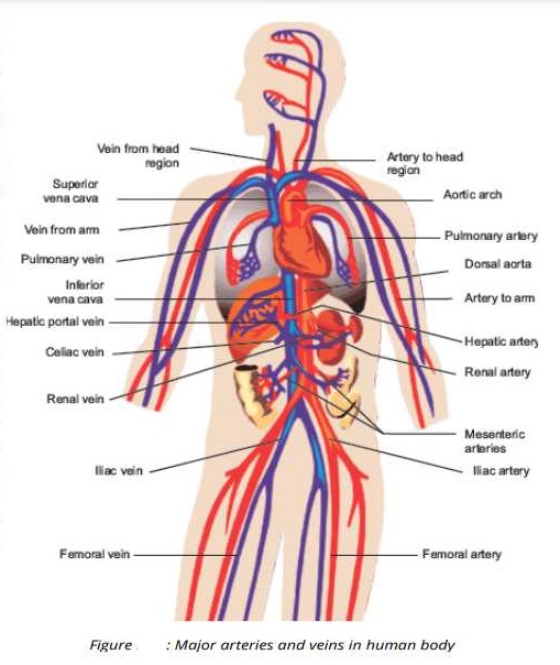
From the right ventricle the pulmonary trunk originates which is further divided into right pulmonary and left pulmonary arteries. These arteries carry the deoxygenated blood to the right and left lungs respectively where it is oxygenated.
The oxygenated blood from the left ventricle of the heart is carried in a large artery called aorta. The aorta then forms an Arch called Aortic arch. Three arteries arise from the upper surface Aortic arch which supply blood to the head, arms and shoulders.
Then the Aorta descends through the chest cavity and passes on to the abdomen and is now called Dorsal Aorta, The dorsal aorta is divided into many branches. Following are the important branches:
1. Intercostal Arteries: These are several in number and supply blood to the Ribs.
2. Coeliac Artery: It supplies blood to liver, stomach and spleen.
3. Superior Mesenteric: It supplies blood to Duodenum, Pancreas and intestine.
4. Hepatic Artery: It supplies blood to the liver.
5. Renal Arteries. These supply blood to the kidneys.
6. Comodal Arteries: These supply blood to the gonads (testes or ovasn)
7. Inferior Mesenteric Artery: This lies below the gonadal arteries and supplies blood to a part of the large intestine and rectum.
In the lower abdominal region, the Aorta bifurcates into common Iliac Arteries. Each common iliac artery divides into an internal iliac Artery and an external Iliac Artery. Each external iliac artery becomes femoral artery in the upper thigh. Femoral artery gives branches to thigh, knee, shank, ankle and foot.
Q.13. How would you differentiate between atherosclerosis and arteriosclerosis?
Answer:
The oxygenated blood is returned to the left atrium from the lungs by right and left pulmonary veins.
Following two major veins carry the deoxygenated blood from the rest of the body and pour into right atrium.
(i) Superior vena cava
(ii) Inferior vena cava
(i) Superior vena cava:
Superior vena cava is formed by the union of different veins (right and left internal juglar veins, right and left external juglar veins and right and left suhclavian veins) from head, shoulders and arms.
(ii) Inferior Vena Cava:
Inferior vena cava is formed by the union of right and left common iliac veins.
Many veins carry the deoxygenated blood back to the atrium of the heart from the legs. Following veins are involved.
1. Femoral Vein:
It is formed by the union of veins that carry blood from calf foot and knee.
2. External Veins:
These veins collect the blood from the hind limbs.
3. A pair of Renal Veins:
These veins collect blood from the kidneys.
4. Hepatic Vein
It collects blood from the liver.
5 Gonadal Veins:
These collect blood from the gonads, i.e. testes and veins blood from the digestive tract (stomach, spleen, pancreas and intestine) and carries blood to the liver.
In the chest cavity (Thoracic cavity), inferior vena cava also receives veins from thoracic walls and ribs.
Q.14. State the causes, treatments and prevention of myocardial infarction.
Answer:
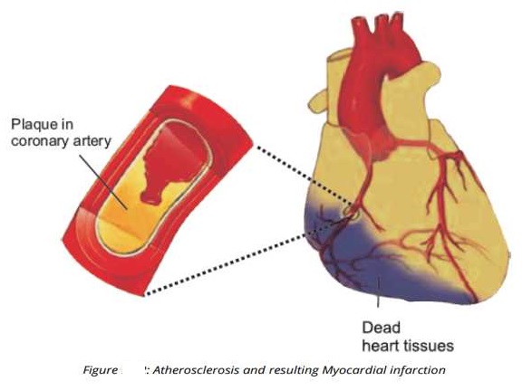
The term myocardial infarction is derived form myocardium (the heart muscle) and infarction (tissue death).
Blockage of blood vessels in the heart by an embolus (or by locally formed thrombus) causes necrosis of a portion of heart muscles, a condition familiarly known as a heart attack or technically myocardial infarction.
Causes and Symptoms
Following are the causes for heart attack:
(i) Blood clotting in coronary arteries.
(ii) Sever chest pain.
(iii) Sensation of tightness, pressure or squeezing.
(iv) Pain radiates most often to the left arm.
(v) Pain may also radiates to the lower jaw, neck, right arm and back.
(vi) Loss of prevention.
(vii) Sudden death.
PREVENTION
We can prevent or avoid these situations or malformation/functions.
(i) Avoid too much fatty food (especially rich in cholesterol).
(ii) Maintain normal body weight.
(iii) Avoid stress and tension.
(iv) Control our blood pressure by regular walk and exercise.
(v) Do not smoke.
TREATMENT
. Treatment with oxygen supply, aspirin and sublingual tablet of glycerol trinitrate.
. Angioplasty (mechanical widening of a narrowed or totally obstructed blood vessel)
. Bypass Surgery (Surgery in which arteries or veins from elsewhere in the patient’s body are grafted to the coronary arteries to improve the blood supply heart muscles).
Cardiovascular disorders are the cause of 12% of adult deaths in Pakistan (Source: Federal Bureau of Statistics of Pakistan). Hypertension (blood pressure higher than normal) is the most common cause of cardiovascular disorders in Pakistan.
. There are over 12 million hypertension patients in Pakistan.
. About 10% of our population is diabetic.
. According to the World Health Organization in Pakistan 1 in 7 urban adults is obese.
SHORT QUESTIONS – TEXT EXERCISE
Q.1. What are lenticels and where are they fund in plant body?
Answer:
Transpiration is the loss of water from plant surface through evaporation. This loss may occur through special openings called lenticels present in the stems of some plants.
Q.2. What is the role of potassium ions in the opening of stomata?
Answer:
The blue wavelengths of daylight cause the K+ to flow into the guard cells, from the surrounding epidermal cells. Water passively follows these ions into the guard cells. The guard cells become turgid and open.
Q.3. Define the cohesion-tension theory
Answer:
Cohesion-tension theory:
According to this theory, the force which carries water (and dissolved materials) upward through the xylem is
transpirational pull. Transpiration creates a pressure difference that pulls water and salts up from roots.
Q.4. What do you mean by sources and sinks according to the pressure flow mechanism?
Answer:
Movement of food in plants has been studied of years. The currently accepted hypothesis states that transport of food is through pressure- flow mechanism. In pressure-flow mechanism, food is moved from sources to sinks. Sources include the exporting organs, typically a mature leaf or storage organ.. Sinks are the areas of active metabolism or storage e.g. roots. Tubers, developing fruits and leaves and growing regions.
Q.5. What are the two main types of white blood cell? How do they differ?
Answer:
White blood cells are divided into two main types on the basis of granules. These are:
(i) Granulocytes (ii) Agranulocytes
1. Granulocytes
• Granulocytes have granules in their cytoplasm and their nucleus is characteristically 2-4 lobed.
• Granulocytes constitute about 71 to 72% of total white blood cell count.
• Granulocytes are formed in red bone marrow.
• Granulocytes include Neutrophils, Eosinophils and Basophils.
2. Agranulocytes
• These have clear cytoplasm (no granules)
• Their nuclei do not have lobed appearance, but are rounded.
• They are about 28 – 29% of total white blood cell count in normal persons.
• Agranulocytes are formed in lymphoid tissue, such as those of the lymph nodes, spleen, tonsils, adenoids and the thymus.
• They have two types: Monocytes and Lymphocytes.
Q.6. You see pus a the site of infection on you skin. How is it formed?
Answer:
WBCs die in the process of killing the germs. These dead cells accumulate and make the white substance called pus, seen-at infection sites.
Q.7. What role does the pericardial fluid play?
Answer:
Heart is enclosed in a sac known as pericardium. These is a fluid, known as pericardial fluid, between pericardium and heart, during heart contractions.
Q.8. Define the terms systole and diastole.
Answer:
Contraction of the heart chambers is called systole while’ relaxation is called diastole. In one heartbeat, diastole lasts about 0.4 see, atrial systole takes about 0.1 see, and the ventricular systole lasts about 0.3 sec.
THE TERMS TO KNOW
