Unit 4 – Cells and Tissues (Exercise Questions)
Answer the following questions.
Q.1. State the cell theory.
Q.2. Explain the functions of cell membrane.
Q.3. Describe the structure of cell wall.
Q.4. Discuss nucleus structure and function.
Q.5. Describe the structure and function of endoplasmic reticulum and Golgi apparatus.
Q.6. Describe the formation and function of lysosomes.
Q.7. Explain what would happen when a plant and an animal cell is placed in a hypertonic solution.
Q.8. Describe the internal structure of chloroplast and compare it with that of mitochondrion.
Q.9. Explain the phenomenon involved in the passage of matter across cell membrane.
Q.10. Describe how turgor pressure develops in a plant cell.
Q.11. State the relation between cell function and cell structure.
Q.12. Describe the differences in prokaryotic and eukaryotic cells.
Q.13. Explain how surface area to volume ratio limits cell size.
Q.14. Describe the major animal tissues (epithelial, connective, muscular and nervous) in terms of their cell specificities, location and functions.
Short Questions-Text Exercise
Q.1. State the cell theory.
Q.2. What are the functions of leucoplasts and chromoplasts?
Q.3. Differentiate between diffusion and facilitated diffusion?
Q.4. What is meant by hypertonic and hypotonic solution?
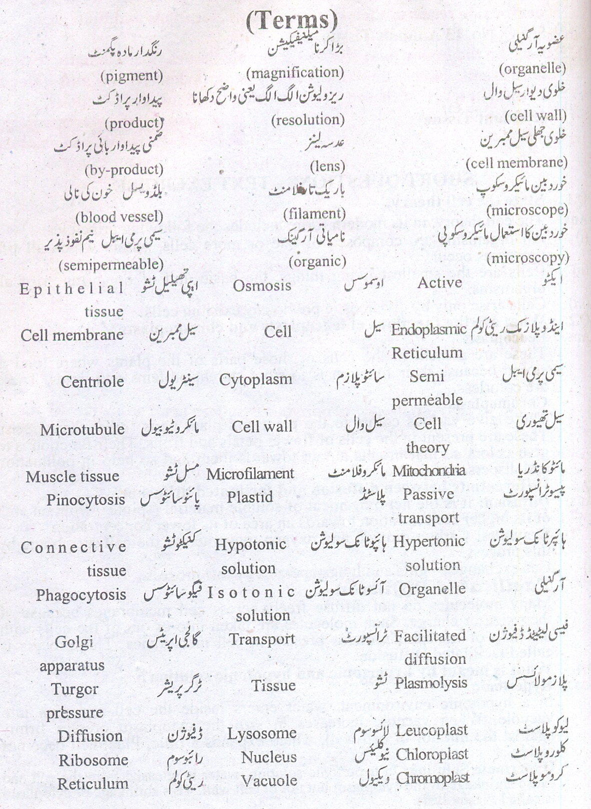
THE TERMS TO KNOW
Q.1. State the cell theory.
Answer:
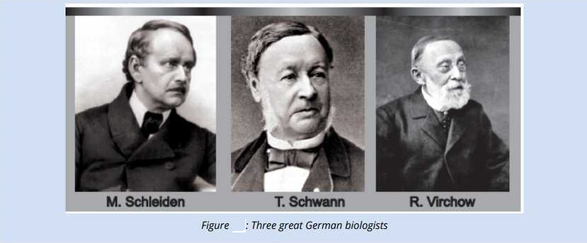
Contributions of M. Schleiden and T. Schwann laid to the formulation of Theory which is called as “Cell Theory”. Their proposed theory can be defined as “All living things are composed of living cells.”
Extension of Cell Theory:
In 1855, Cell Theory was extended by the idea of a German Physician, Rudolf Virchow. He proposed that all living cells arise from pre-existing cells (“Omnis cellula e celula”). This idea was experimentally proved by Louis Pasteur in 1862.
Salient Features:
The salient features of the most modern cell theory are as follows:
(i) All animals and plants are made of cells and cell products. Among these some organisms are unicellular and
some are multicellar.
(ii) Cells are structural and functional units of living organisms.
(iii) New cells come from the pre-existing cells by cell division.
Q.2. Explain the functions of cell membrane.
Answer:
Cell Membrane:
Plasma membrane or cell membrane is the outer most boundary of the animal cells but in plant cells, it is covered by a cell wall.
Chemical Composition:
Cell membrane is chemically composed of:
a. 60 – 80 % Protein.
b. 20 – 40 % Lipids
In addition there is a small quantity (2-10%) of carbohydrates.
Structure:
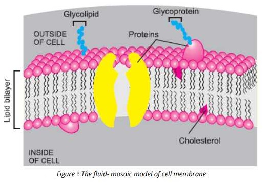
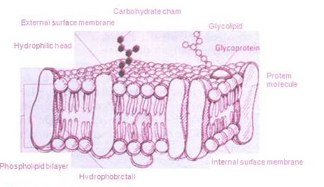
Structurally, cell membrane is composed of layers of different materials and has extremely small pores in it through which only small molecules can pass. There are different theories about the ‘structure of plasma membrane. i. In 1935 Danielli and Davson proposed that cell membrane consists of three layers.
ii. In 1938 Harvey and Danielli suggested that plasma membrane is composed of lipid bilayer sandwitched between
two protein layers. This basic structure is found in all the membranes such as those of, mitochondria;
chloroplasts etc, and are called unit membrane.
Q.3. Describe the structure of cell wall.
Answer:
Cell Wall:
It is the outer semirigid and non-Living boundary of a plant cell.
Chemical Composition:
In its chemical composition cellulose is the most prominent substance, where, the cell wall of fungi is made up of chitin. .
Structure:
Thickness of a cell wall varies in different cells e.g in plants, xylem, vessels elements and tracheids have thick walls while parenchyma cells have thin walls.
The primary layer of the cell wall is called primary wall, which is further strengthened by an additional layer called secondary wall, especially in xylem vessels. It is comparatively thicker than primary wall. The cell walls of neighbouring cells are connected by cytoplasmic bridges which are called Plasmodesmata. Through these connections, cells transfer chemicals among each other.
Functions:
(i) Shape & rigidity: Cell wall provides a definite shape to the cell and keeps it rigid.
(ii) Permeable: It does not act, as a barrier to the materials passing through it.
(iii) Support & Strength: Cell wall provides mechanical strength and skeletal support for the
individual cell and to the plant as a whole.
(iv) Movement of water & minerals: Water and minerals can. move through an interconnected
system of cell wall (i.e the apoplast).
Protection: Cell wall protects the cells from osmotic lysis.
Q.4. Discuss nucleus structure and function.
Answer:
Nucleus:
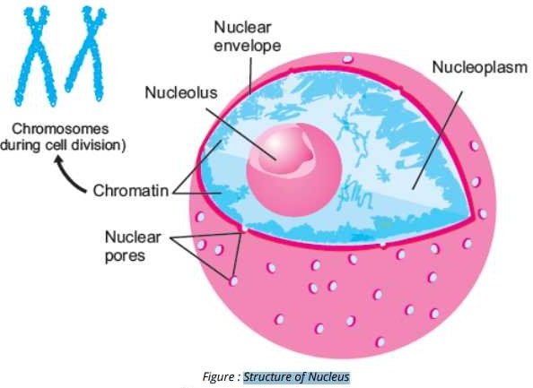
The most important and visible part of a cell is nucleus. It is present in the center of the animal cell but due to presence of large central vacuole, it is pushed to one side in the plant cell.
Structure
Its structure contains various component parts e.g.
Nuclear Membrane:
The outer most covering of nucleus is called Nuclear membrane. It is a double membrane and. also pores are present in it.
Nucleoplasm:
The granular matrix present inside cell is called Nucleoplasm.
Nucleoli:
In the Nucleoplasm, one or more nucleoli are present. These are usually visible as dark spots and are responsible for the formation of ribosomal RNA.
Chromosomes:
Chromosomes or genetic material is also present in nucleoplasm in the form of chromatin (thread like structure).
Chromosomes are made up of Deoxyribonucleic acid (DNA) and histone proteins.
DNA contains the message for the synthesis of cellular proteins. This message is read by messenger RNA which carries it to ribosome for the protein synthesis.
Functions
It controls all the activities of the cell and is also responsible for the transmission of characteristics to the next generation.
Q.5. Describe the structure and function of endoplasmic reticulum and Golgi apparatus.
Answer:
Endoplasmic Reticulum (ER):
This is a complex membranous system, which consists of two unit- membranes enclosing a narrow space about 4mm wide. These form a network of interconnected channels that extends from cell membrane to the nuclear: membrane.
Types
There-are two types of ER.
(i) Rough Endoplasmic Reticulum.
(ii) Smooth Endoplasmic Reticulum.
Rough Endoplasmic Reticulum:
Numerous Ribosomes are present on this type of Endoplasmic Reticulum
giving it a rough texture.
Smooth . Endoplasmic Reticulum:
These are free from Ribosomes.
Functions:
(a) Rough Endoplasmic Reticulum helps in protein synthesis.
(b) Smooth Endoplasmic Reticulum is involved in lipid metabolism.
(c) These also. help in transport of materials from one part of the cell to the other part. .
(d) These also help to detoxify harmful chemicals that have entered the cell.
Golgi Apparatus:
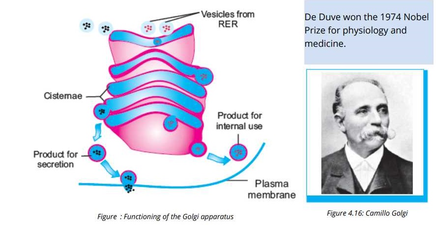
Golgi apparatus or Golgi complex is an assemblage of flat or curved sacs (cisternae) lying one above the-other.
Discovery:
These were discovered by Camillo Golgi” in 1898 in the neural tissue of the animals.
Functions:
(i) These are concerned with cell secretions, which are products formed within the cell on Ribosomes and then
passed to the outside through endoplasmic reticulum and Golgi apparatus. These secretions are converted
into finished product.
(ii) These also modify the proteins and lipids by adding carbohydrates and convert them into glycoproteins or
glycolipids.
(iii) These are also involved in lysosome synthesis.
Q.6. Describe the formation and function of lysosomes.
Answer:
Lysosomes:
Lysosomes are cytoplasmic organelles and are different from others due to their morphology. Lysosomes (Lyso = splitting; soma = body) are found in most eukaryotic cells. They are most abundant in those animal cells, which
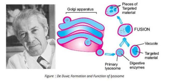
exhibit phagocytic activity. They are bound by a single membrane and are simple sacs rich in acid phosphatase and several other hydrolytic enzymes.
These enzymes are synthesized on RER and are further processed in the Golgi apparatus. The processed’ enzymes are budded off as Golgi vesicles and are called as primary Lysosomes.
 Discovery:
These were discovered in the -mid-twentieth century by a Belgian Scientist “de Duve”.
Functions:
Because lysosomes contain strong digestive enzymes so these work for the break-down of food and waste materials inside the cell.
Q.7. Explain what would happen when a plant and an animal cell is placed in a hypertonic solution.
Answer:
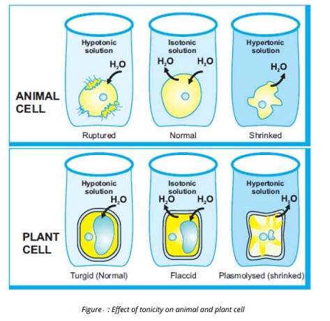
Environment can never be same as the environment inside the cell. So both animal and plant cells have a lot of problems to maintain the water quantity, When an animal cell e.g blood cell is placed in an isotonic solution then no change in shape of all takes place because the water moving outside in cell becomes balance ‘with water moving inside. But when cell is placed in a hypertonic solution (whose concentration is higher than inside of cell) then
water of the cell will move outside and cause the shrinkage of cell.
When cell is placed in a Hypotonic solution (whose solute concentration is lower than inside of cell) then water will enter into the cell and cause the bursting.
So cells of animals in freshwater (hypotonic) and in seawater (hypertonic) must have some ways to maintain the water quantity of cell.
Q.8. Describe the internal structure of chloroplast and compare it with that of mitochondrion.
Answer:
Chloroplasts:
These are present in the green parts of the plant particularly in leaves.
Structure of Chloroplasts:
Each chloroplast is bounded-by a double membrane. The outer membrane is smooth. Inner membrane gives rise to stacked membrane system composed of sacs which are called thylakoids. Stacks of thylakoids are called Grana.
Chloroplasts are filled with semi fluid matrix called stroma. Thylakoids contain green pigments (chlorophyll).
Function of Chloroplast
Due to the chlorophyll, photosynthesis takes place in them.
Q.9. Explain the phenomenon involved in the passage of matter across cell membrane.
Answer:
Cell membranes maintain balance inside the cell as well as outside. This balance is. maintained by various phenomena. Which are arranged in the following:
TRANSPORT OF MATERIALS
Non-Facilitated Transport:
Non-polar molecules like oil droplets, phospholipids, fatty acids etc. move across the membrane freely through the lipid bilayer. This is called non- facilitated transport.
Facilitated Transport
The movement of ionic material like water molecules, O2, CO2, ions or radicals is carried out across the cell membrane only with the help of proteins. So it is called facilitated transport.
There are two types of facilitated transport:
(i) Facilitated Active Transport,
(ii) Facilitated Passive Transport,
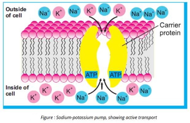
(i) Facilitated Active Transport:
The transport of molecules across the membrane against the concentration gradient (from lower to higher concentration) with the expenditure of energy is called Facilitated Active Transport. The required energy is in the
form of ATP?
Example
Membrane’s of nerve cells have some earner proteins e.g. Sodium- Potassium Pump”.
In a resting (not conducting nerve impulse) nerve cell. This pump uses energy in the form of ATP to maintain higher concentrations of K+ and lower concentrations of Na+ inside the cell. For this purpose the pump actively moves NA+ to the outside of the cell where they are already in higher concentration. Similarly this pumps moves K+ from outside to inside the cell where they are in higher concentration.
(ii) Facilitated Passive Transport:
The transport of molecules along the concentration gradient (from higher to lower concentration) without utilizing energy is called facilitated passive transport.
There are two types of Facilitated Passive Transport:
(a) Diffusion (b) Osmosis
(a) Diffusion:
It is the net movement- of soluble material (solutes) from an area of its higher concentration towards an area of its lower concentration.
Examples
(i) Carbon dioxide and oxygen move across the cell membrane by this process.
(ii) Gas exchange in gills and lungs operates by this process.
Facilitated Diffusion
Many molecules do not diffuse freely across cell membranes because of their size or charge. Such molecules are taken into or out of the cells with the help of transport proteins present in cell membranes. This process is
called facilitated Diffusion.
Osmosis:
The movement of water molecules” (solvent) from an area of its higher concentration to an area of its lower concentration through a selectively I semi permeable membrane is called Osmosis.
Example
The rule of osmosis can be best understood through the concept of tonicity of the solutions. The term tonicity refers to the relative concentration of solutes in the solutions being compared.
• Hypertonic solutions are those in which more solute is present.
• Hypotonic solutions are those with less solute.
• Isotonic solutions have equal concentrations of solutes.
In hypertonic solution the solute molecules attract clusters of water molecules, so the fewer water molecules are free to diffuse across the membrane. On the other hand in a hypertonic solution with fewer. solute molecules, there are more free water molecules and there is net movement of water from hypotonic solution to the hypertonic solution.
Filtration
This is the process in which small molecules are forced to move across the semi permeable membrane with the help of Hydrostatic (water) pressure or blood pressure.
For example in the body of an animal, blood pressure forces water and dissolved, molecules – to move through the semi-permeable membranes of the capillary” wall cells. In filtration -the pressure cannot force large molecules such as proteins, to pass through the membrane pores.
Endocytosis
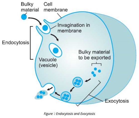
In this process, bulky materials are moved to inside from outside of the cell. The various steps involved in this process are as follows:
(i) When a useful bulk material come in proximity of cell then cell membrane invaginates.
(ii) The bulky material is captured inside this invagination.
(iii) Then two ends of membranes are sealed together and a small vesicle is formed which may be a food vacuole.
(iv) Food vacuole or vesicle detaches and freely move in cytoplasm.
Types
There are two types of Endocytosist.
(i) Phagocytosis:
When Endocytosis of a solid material in the form of droplets takes place then this is called Phagocytosis also called cellular eating.
(ii) Pinocytosis:
When Endocytosis of a liquid material takes place then this IS called Pinocytosis also called cellular drinking.
Exocytosis:
The process in which bulky material is expelled to the outside of cell is called Exocytosis.
The various steps of this process is as follows:
(i) Waste material is packed inside a vesicle.
(ii) Vesicle moves to the cell membrane.
(iii) Vesicle fuses to the membrane and releases its contents into the extracellular environment.
This process adds new membrane which replaces the part of cell membrane lost during endocytosis.
Q.10. Describe how turgor pressure develops in a plant cell.
Answer:
Water balance problems are somewhat different for plant cells because of the presence of rigid cell walls. In a hypotonic environment, water enters inside the cell and then into vacuole. When vacuole increases in size the’
cytoplasm presses firmly against the interior of cell wall, which expands a little. Plant cell becomes rigid. The internal pressure of such a rigid cell is known as Turgor Pressure and process is called Turgor.
In isotonic environment the plant cell is flaccid (loose / not firm), because the net uptake of water is not enough to make it turgid.
In hypertonic solution, water is released from the cell and cause shrinkage of the cytoplasm but not of cell wall. This shrinkage of cytoplasm is called Plasmolysis.
Q.11. State the relation between cell function and cell structure.
Answer:
RELATIONSHIP
The bodies of animals and plants are made up of different cell types. Each type performs specific function and all coordinated functions perform the life processes of organism.
Types of Cells:
Human body is made up of about 200 types of cells. Cells of one type may differ from those of other types in the following respects:
Size and Shape:
• Red blood cells are round to accommodate globular haemoglobin
• Nerve cells are long for the transmission of nerve-impulses.
• Xylem cells are tube-like and have thick walls for conduction of water and support.
Surface-Area to Volume Ratio:
• Root hair cells have large surface area for maximum absorption of water and salts.
Presence or absence of organelles:
• Cells involved in making secretions have more complex ER and Golgi apparatus.
• Cells involved in photosynthesis have chloroplasts.
Functions of cells:
Individual cells contribute to the functioning of the whole body. It can be explained by the
following examples of human body cells:
Nerve Cells:
Nerve cells conduct nerve conduct impulses and thus contribute to the coordination in body.
Muscle Cells:
Muscle cells undergo contraction and share their role in movements in body.
Red Blood Cells:
Red blood cells carry oxygen and so contribute in the role of blood in transportation.
White Blood Cells:
White blood cells kill foreign agents and so contribute in the role of blood in defence.
Skin Cells:
Some skin cells act as physical barriers against foreign materials and some act as receptors
for temperature, touch and pain.
Bone Cells:
The cells of bone deposit calcium in their extracellular spaces to make the bone tough and
thus contribute to the supporting role of bones.
Cells as an Open System:
A cell works as an ‘open system’. i.e. it takes in substances needed for its metabolic activities
through its cell membrane. Then it performs the metabolic processes assigned to it. Products
and by-products are formed in metabolism. Cell either utilizes the products or transports
them to other cells. The by-products are either stored or are excreted out of the cell.
Q.12. Describe the differences in prokaryotic and eukaryotic cells.
Answer:
Cells are basically of two types:
a. Prokaryotic Cells.
b. Eukaryotic Cells.
a. Prokaryotic Cells:
These are the simpler cells which do not have membrane-bounded nucleus and various other organelles.
Eukaryotic Cells:
All the plants and animals are made up of Eukaryotic cells which have membrane-bounded nuclei and various other organelles are also present.
Differentiation among them:
| Prokaryotic Cells | Eukaryotic Cells |
| 1. The organisms made of prokaryotic cells are called Prokaryotes e.g bacteria and cyanobacteria. | 1. The organisms made of eukaryotic cells are called eukaryotes e.g animals plants fungi and protests. |
| 2. Chromosomes are present in the cytoplasm and no membrane bounded nucleus is resent. | 2. Chromosomes are present in membrane bounded nucleus. |
| 3. No membrane bounded cell organelles are resent. | 3. Membrane bounded organelles are present. |
| 4. Ribosomes, are of small size and freely scattered in cytoplasm. | 4. Ribosomes are of large size and are present on endoplasmic reticulum or free in cytoplasm. |
| 5. Cell wall is composed of peptidoglycan or murein. Cellulose is absent. | 5. Cell wall of plant cell is composed of cellulose while in fungi it is of chitin. |
| 6.These cells are simple, comparatively smaller in size (average diameter 05-10 µm). | 6. These cells are complex, comparatively larger in Size (average diameter 10-100µm). |
Q.13. Explain how surface area to volume ratio limits cell size.
Answer:
Cell sizes are directly related to their function. This idea can be explained with the help of examples, e.g.
(i) Bird’s eggs are the bulkiest cells. These are bulky because these contain a large ‘amount of nutrients for
developing young.
(ii) An animal’s body, muscle cells and nerve cells are the longest. Long muscle cells arc efficient in pulling
different body parts together. The benefit of lengthy nerve cells is that these can transmit nerve signals
between distant parts of animal’s body.
(iii) Human Red blood cells are very small just because these could easily travel through Sum diameter of tiniest
blood vessels.
Similarly, surface area is related to the size of cell. Surface area is just the area of the boundary of total volume. Volume is the total length, width and height of cells. Small cells have high surface area and large cells have low surface area as compared to their Volumes.
All the activities of the cell depend upon the availability of nutrients and removal of wastes. Both these functions are performed through the cell surface membranes. So there must be a large surface area as compared to
the volume of cells.
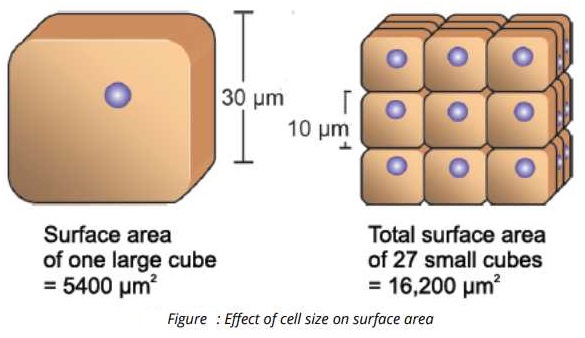
Most cells are small in size: In relation of their volumes, large cells have less surface areas as compared to small cells. Figure 4.21 shows this relationship using cube-shaped cells. The figure shows, 1 large cell and 27
small cells. In both cases, the total volume is same:
Volume = 30 µm x 30 µm x 30 µm = 27,000 µm3
In contrast to the total volume, the total surface areas are very different. Because a cubical shape has 6 sides, its surface area is 6 times the area of 1 side, The surface areas of cubes are as follows:
Surface area of 1 large cube = 6 X (30µn X 30µm) = 5400µm2
Surface area of 1 small cube = 6 x ( 10µm X 10µm) = 600 µm- and
Surface area of 27 small cubes = 27 x 600 µm2 = 16,200 µm2
Q.14. Describe the major animal tissues (epithelial, connective, muscular and nervous) in terms of their cell specificities, location and functions.
Answer:
Animal Tissues:
Four types of tissues are found in animal body e.g.
1. Epithelial Tissue.
2. Connective Tissue.
3. Muscle Tissue.
4. Nerves Tissue.
Epithelial Tissue:
These tissues cover the outside of the body and lines organs and cavities.
Properties
(i) The cells in this tissue are closely packed with very little space among them.
(ii) These help to protect organisms from microorganisms, injury fluid loss.
Types
There are various types of epithelial tissue on tile basis of shape as well as the number of cell layers. These types are as follows:
(i) Simple Squamous Epithelium:
These cells are arranged in fine tightly packed layer and form the outer surface of skin of organisms. They control the diffusion and filtration of the substances.
(ii) Simple Cuboidal Epithelium:
These consist of single layer of tightly packed cells on the inner surface of tubular organs. These are cuboids in shape.
These make secretions and absorb materials.
(iii) Simple Columnar Epithelium:
These consist of single layer of elongate cells These are found in the lining of digestive tract and gallbladder etc.
These secrete enzymes.
(iv) Ciliated Columnar Epithelium:
In this category, each columnar cell contains a tuft of cilia at the top. These are found in the lining of trachea and bronchi.
These cells propel mucous by ciliary action.
(v) Stratified Squamous Epithelium:
These are composed of many layers of flattened cells. These are found in the inner lining of esophagus, mouth and at the surface of the skin.
These protect underlying tissues from abrasion.
Short Questions-Text Exercise
Q.1 State the cell theory.
Answer:
The cell. theory, in its modem form, includes the following principles:
(i) All organisms are composed of one or more cells, within which all life processes occur.
(ii) Cells are the smallest living things, the basic unit of organization of all organisms.
(iii) Cells arise only by divisions in previously existing cells.
Q.2. What are the functions of leucoplasts and chromoplasts?
Answer:
Leucoplasts
These are present .in the cells or those parts of the plants where food is stored because their function IS to store starch, proteins and lipids. These are colorless.
Chromoplasts
These give various colors to the plant and plant parts (other than green). These are present In. the cells of flower petals and fruits. Their function is to give colors. so that Insects attract towards them and so help in pollination and dispersal of fruits.
Q.3. Differentiate between diffusion and facilitated diffusion?
Answer:
Diffusion: It is the net movement of soluble material (solutes) from an area of its higher concentration towards an area of its lower concentration.
Examples: Carbon dioxide and oxygen move across the cell membrane b this process.
Gas exchange in gills and lungs operates by this process.
Facilitated Diffusion
Many molecules do not diffuse freely across’ cell membranes because of their size or charge. Such molecules are taken into or out of the cells with the help of transport ‘proteins present in cell membranes. This process is
called facilitated Diffusion.
Q.4. What is meant by hypertonic and hypotonic solution?
Hypertonic
In a’ hypotonic environment, water enters inside the cell and then into vacuole. When vacuole increases in size the cytoplasm presses firmly against the interior of cell wall, which expands a little. Plant cell becomes rigid.
Hypertonic Solution: In hypertonic solution, water is released from the cell and
cause shrinkage of the cytoplasm hut not of cell wall. This shrinkage of cytoplasm is called Plasmolysis.
THE TERMS TO KNOW
