Q.5 Explain Central Nervous System (C.N.S) in detail. Parts of CNS
CNS consists of two parts:
(i) Brain” (ii) Spinal cord
(i) Brain
Introduction
In animals, all life activities are under the control of brain. The structure of brain is suitable to perform this function. ”
Location
Brain is situated inside a bony cranium i.e. a part of skull.
Meninges
Inside cranium, brain is covered by three layers called meninges.
Function of meninges
Meninges protect brain and also provide nutrients and oxygen to brain tissues through their capillaries. Ventricles
The brain contains fluid filled ventricles that are continuous with the central canal of spinal cord.Cerebrospinal fluid (CSF)
Fluid within ventricles and central canal is called cerebrospinal fluid (CSF).
Function of CSF
It provides cushioning and ions to brain and spinal cord.
Division of Brain
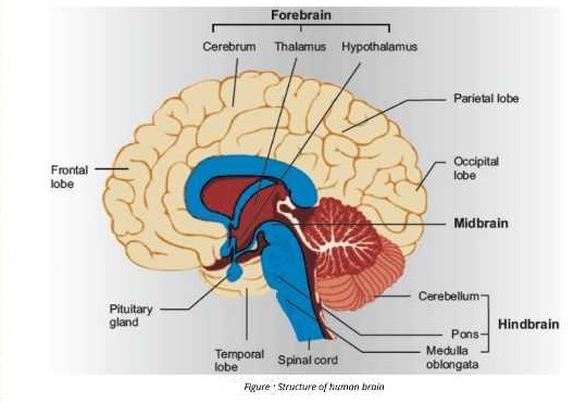
There are three main regions in the brain of humans and other vertebrates. These are forebrain, midbrain, and hindbrain.
(a) Forebrain
Introduction
Forebrain is the largest area of brain. It is most highly developed in humans.
Parts of Forebrain
(i) Thalamus
Location
It lies just below the cerebrum.
Functions
It serves as a relay center between various parts of brain and spinal cord. It also receives and modifies sensory impulses (except from nose) before they travel to cerebrum. Thalamus is also involved in pain perception and consciousness i.e. sleep and awakening.
(ii) Hypothalamus
Location
It lies above midbrain and just below thalamus.
Size
In humans, it is about the size of an almond.
Function
(a) The most important function of hypothalamus is to link nervous system and endocrine
system.
(b) It controls the secretions of pituitary gland.
(c) It controls feelings such as rage, pain, pleasure and sorrow.
(iii) Cerebrum
It is the largest part of forebrain.
Function
It controls skeletal muscles, thinking, intelligence and emotions.
Cerebral hemispheres
Çerebrum is divided into two cerebral hemispheres.
Olfactory Bulbs
The anterior parts of cerebral hemispheres are called olfactory bulbs which receive impulses from olfactory nerves and create the sensation of smell.
Cerebral cortex
The upper layer of cerebral hemisphere i.e. cerebral cortex consists of grey matter.
Grey matter
The grey matter of nervous system consists of cell bodies and non-myelinated axons.
White matter
Beneath this layer is present the white matter. The white matter of nervous system consists of myelinated axons. Cerebral cortex has a large surface area and is folded in order to fit in skull.
Lobes of cerebral cortex
It is divided into four lobes:
| Lobe | Functions |
| Frontal | Controls motor functions, permits conscious control of skeletal muscles and coordinates movements involved in speech |
| Parietal | Contains sensory areas that receive impulses from skin |
| Occipital | Receives and analyzes visual information |
| Temporal | Concerned with hearing and smell |
Hippocampus
It is a structure that is deep in the cerebrum. It functions for the formation of new memories. People with a damaged hippocampus cannot remember things that occurred after that but can remember things that occurred before damage.
(b) MIDBRAIN
Location:
Midbrain lies between hindbrain and forebrain and it connects the two.
Functions:
1. It receives sensory information and sends it to the appropriate parts of fore brain.
2. Midbrain also controls some auditory reflexes and posture.
(c) HINDBRAIN / What are parts of hind brain and how they perform their function? Parts of hindbrain
Hindbrain consists of three major parts:
1. Medulla Oblongata
Location
It lies on the top of spinal cord.
Functions
(i) It controls breathing, heart rate and blood pressure.
(ii) It also controls many reflexes such as vomiting, coughing, sneezing etc.
Information that passes between spinal cord and the rest of brain pass through medulla.
2. Cerebellum Location
It is behind the medulla oblongata.
Function
It coordinates muscle movements.
3. Pons Location
It is present on the top of medulla oblongata.
Function
(i) It assists medulla in controlling breathing.
(ii) It also serves as a connection between cerebellum and spinal cord.

(ii) SPINAL CORD
Definition
It is a continuation of medulla oblongata. .
Location
It starts from brain stem and extends to lower back
STRUCTURE
The spinal cord is infact a tubular bundle of nerves. It is the continuation of medulla. oblongata.
Length
It is roughly 40 cm long.
Width
It is about as wide as our thumb for most of its length.
Meninges
Like brain, spinal cord is also covered by meninges.
Protection of spinal cord
The vertebral column surrounds and protects spinal cord.
Outer Region
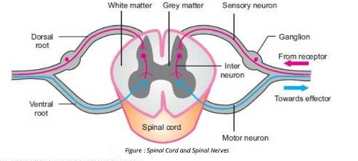
The outer region of spinal cord is made of white matter containing myelinated axons.
Central Region
The central region is butterfly-shaped that surrounds the central canal…. Composition
It is made of grey matter containing neuron cell bodies.
SPINAL NERVES
31 pairs of spinal nerves arise along spinal cord. These are “mixed” nerves because each contains axons of both sensory and motor neurons.
ROOTS OF SPINAL NERVES
At the point where a spinal nerve arises from spinal cord, there are two roots of spinal nerves. Both roots unite and form one mixed spinal nerve.
Dorsal root
The dorsal root contains sensory axons and a ganglion where cell bodies are located.
Ventral root
The ventral root contains axons of motor neurons.

Functions of Spinal Cord
1. It serves as link between body parts and brain. Spinal cord transmits nerve impulses from body parts to brain and from brain to the body parts.
2. Spinal cord also acts as a coordinator, responsible for some simple reflexes.
![]()